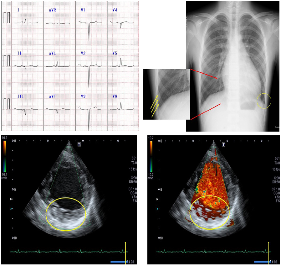
Figure 1: Examination findings at first hospital visit
a) ECG: QS patterns in leads II, III, aVf and V1-4 and T wave inversion in V5 and V6 were found.
b) Chest X-ray: Cardiomegaly (CTR=61%) and lung congestion (dull costophrenic angle, yellow circle; Kerley B-line, yellow arrow) were found.
c) Echocardiogram (short-axis view): Excessive trabeculations and deep recesses are found in the posterior wall of the LV.
d) Echocardiogram (short-axis view): Blood flow in the recessed area on color Doppler echocardiogram.
N/C ratio (ratio of noncompacted layer to compacted layer) was 2.1, compatible with Jenni’s diagnostic criteria [3] for LVNC (N/C >2.0).