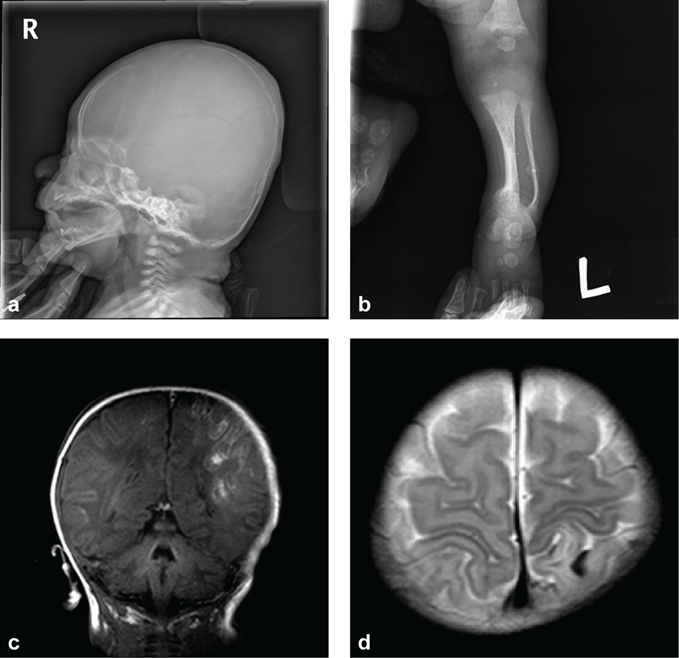
Figure 1: Radiographic findings of the newborn patient with osteogenesis imperfecta. (a) Osteolytic cortical thinning with decreased bone density of skull and flattening of skull base is noted on plain radiograph. (b) The plain radiograph of left knee shows bowing deformity of left tibiofibular shafts and fracture with callus formation of left fibula. Also, there is irregularity and spraying in metaphyseal ends of femur and tibiofibula. (c) In the brain magnetic resonance imaging scan, T1 weighted image shows gyri-form high signal intensity in both parietal lobe(left>right) with some high signal intensity foci in the sulci or subcortical white matter. (d) On T2 weighted image, periventricular and subcortical increased signal intensity on both parietal lobes (left > right) is noted. Additionally, there are multifocal parenchymal and gyral fluid collections with dark signal foci, mainly in left parietal lobe, indicating hemorrhage. This patient also had subdural hemorrhage in posterior fossa (not shown).